April 7, 2017
 I was feeling pretty nervous about the biopsy. The idea of a long needle going into me and then into this mass and pulling something out of it did not sit well. I had a fear that I would feel something with the needle going in and make some movement that was going to cause a serious problem while this needle was in me. They explained to me that I was not going to be fully put under, but I was going to be given something that would make me not feel any of the pain. I would be awake though it, but would most likely not remember all the details of the procedure.
The transport came and it was time to go down to the procedure room. Dr S came into the waiting area and explained the procedure. He is a very good Dr and explained everything quite thoroughly step by step. The biopsy was being done with the aide of the CT scan to help guide the biopsy needle to the correct location. These were the steps as they were explained to me
- I was going to be laying on my stomach on the table and they were going to do an initial scan to identify the entry point on my back.
- The Dr was going to mark the entry point on my back for the needle to go into
- The Dr was going to perform a small incision on my back at the entry point to help get the needle started. The initial needle is a guiding needle with a wide opening that will have a smaller needle inserted through it once it reaches the right location.
- The Dr begins inserting the needle a little bit at a time taking pictures with the CT scan in between each move of the needle to make sure it is heading to the right location.
- Once the guiding needle reaches the biopsy location, the smaller needle is inserted along with a tube that sucks out a piece of the tissue
- The tissue sample is extracted and the needle is removed from my back
Although I never fell asleep it did seem that I lost track of time. I remember a couple of small things during the procure and the table moving to slide me in and out of the CT scan ring, but everything else is just a blur. The next thing I remember is the Dr saying we were all done and my wife being right there as they moved me from the CT scan table to the hospital bed to be transported back.
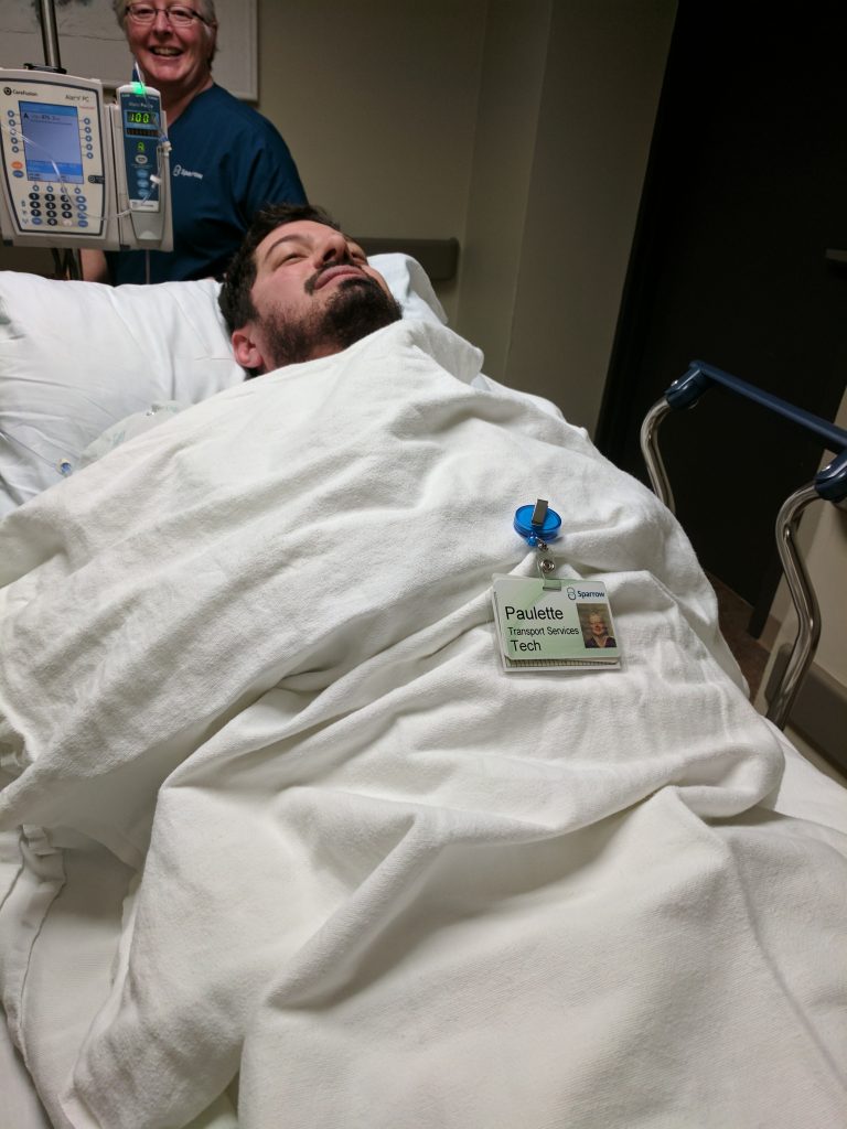
Below is a picture taken of me going back to my room after the procedure. The lady who transported me was named Paulette and let us borrow her name tag for the picture. I was still half out of it at the time.
Below is the actual narrative from Dr S taken from my patient portal
IR retroperitoneal mass biopsy.
HISTORY: Mass. RIGHT groin pain.
TECHNIQUE: CT-guided retroperitoneal mass biopsy.
FINDINGS: Following discussion with the patient and his wife regarding the procedure, its indications, risks, benefits, and possible complications consent was obtained. The patient was brought to the CT suite where he was placed prone on the gantry
table. Continuous physiologic monitoring of the patient’s vital signs was performed throughout the procedure. Conscious sedation was employed utilizing incremental doses of intravenous fentanyl and Versed. These were delivered and monitored by trained
cardiovascular nurse. In total 4 mg of Versed and 100 mcg of fentanyl were utilized for this purpose. Time of sedation 35 minutes.Preliminary axial images again demonstrate the presence of a retroperitoneal mass as well as RIGHT retrocrural lymphadenopathy. A site was selected for percutaneous sampling. The skin was prepped and draped in a routine sterile manner. Following local
anesthesia a 17-gauge guiding needle was advanced into the mass in question. Having verified the needle tip to be appropriately positioned a series of 8 automated core samples were obtained. Feeling these to be adequate the guiding needle was removed.
Postprocedure scans failed to demonstrate evidence of a complicating process. The patient tolerated this well and was transferred to his room in stable condition.